Figure 5
From: Linoleate appears to protect against palmitate-induced inflammation in Huh7 cells

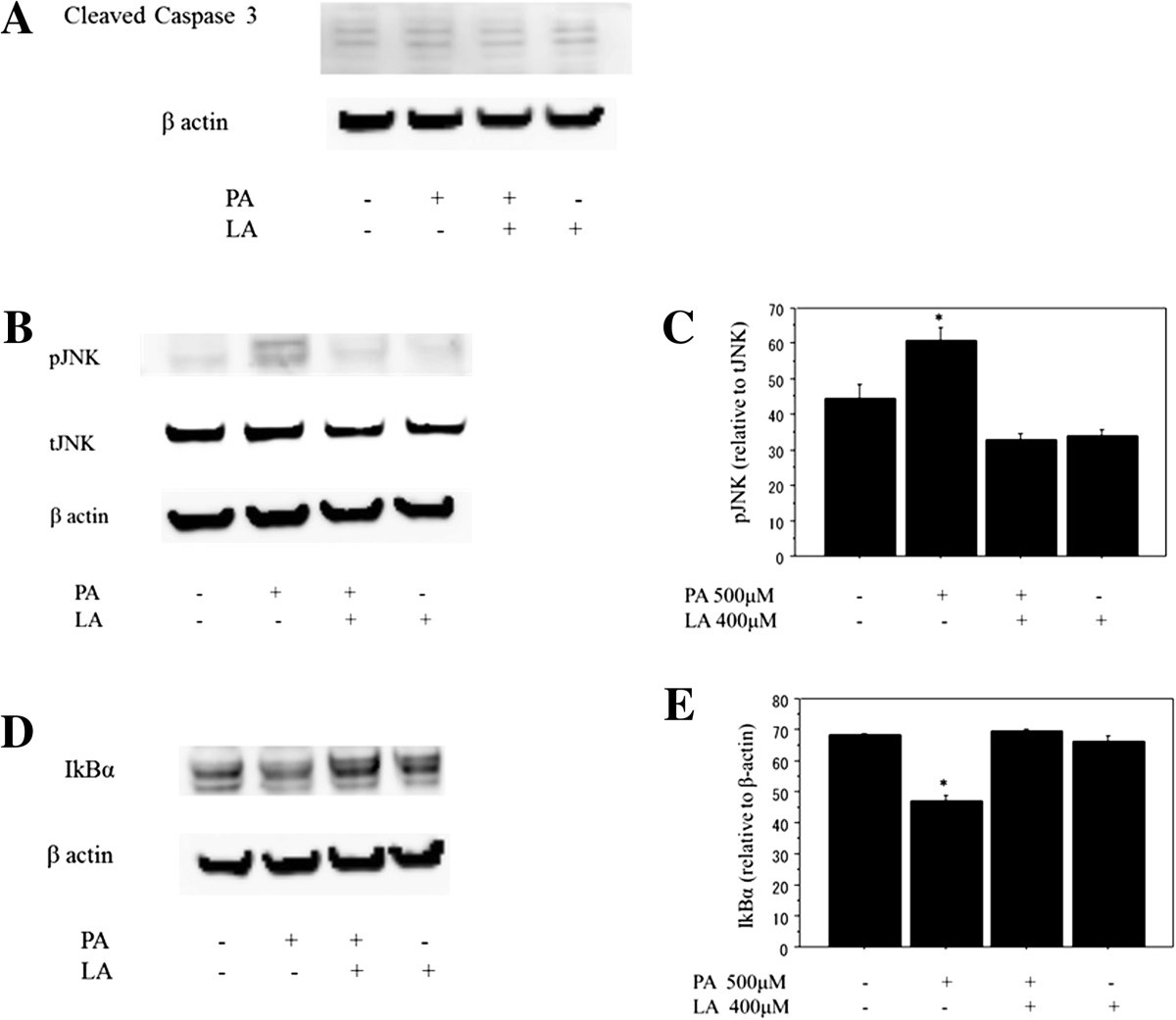
Analysis of protein expression by western blotting. A. Cleaved caspase-3. Caspase-3 was used as an indicator of apoptosis in Huh-7 cells and cleaved caspase-3 was examined after free fatty acid treatment to determine the presence of apoptosis at a molecular level. The cleaved caspase-3 (17 kDa and 19 kDa) showed no significant changes in the expression after the FFA treatment (n = 4), among cells treated by 500 μM palmitate, 400 μM linoleate and co-incubation with both of them. B, C, D, E. The effect of co-incubation with palmitate and linoleate on markers of inflammation. Huh7 cells were incubated with bovine serum albumin (control) or after free fatty acid treatment (500 μM palmitate and/or 400 μM linoleate). Western blot analysis of phospho-c-Jun N-terminal kinase and total-c-Jun N-terminal kinase or β-actin (Figure 5B, 6 hr incubation), IkBα (Figure 5D, 3 hr incubation) and β-actin (internal control). The gels shown are representative of 4 independent experiments and data in the graphs are expressed as the ratio of the target protein to total-c-Jun N-terminal kinase or β-actin (Figure 5C and 5E). The expression of pJNK increased significantly in Huh-7 cells treated with 500 μM palmitate (64.9 ± 4.1) compared to control cells (35 ± 4.2; *p<0.05), cells treated with both 500 μM palmitate and 400 μM linoleate (32.3 ± 2.5; *p<0.01), and cells treated with 400 μM linoleate (36.1 ± 3.3; *p<0.01; n = 4). The expression of IkBα decreased significantly in Huh-7 cells treated with 500 μM palmitate (46.6 ± 2.2) compared to control cells (68.1 ± 5.9 ; *p<0.05), cells treated with both 500 μM palmitate and 400 μM linoleate (68.3 ± 5.7; *p<0.05), and cells treated with 400 μM linoleate (65.7 ± 3.3; *p<0.05; n = 4).
